Exceptional Surgery
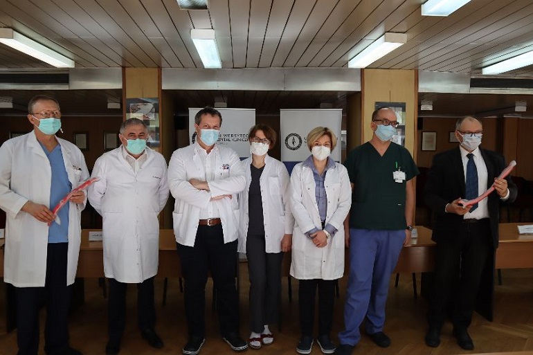
An interdisciplinary team of doctors from Opole and Wrocław, with the support of engineers from the Opole University of Technology, carried out a complex surgery to remove an extensive tumour, reaching from the reproductive organs to the heart. So far only a few such cases have been reported worldwide.
“We admitted the patient to the ward because in the ultrasound examination we found the presence of a large, abnormal structure inside the heart, which extended far down inside the inferior vena cava, as far as the height of the pelvis. The initial diagnosis on the CT scan indicated a thrombus, especially as the patient had suffered a pulmonary embolism a year earlier”. - says Prof. Marek Gierlotka, Head of the Cardiology Ward at the University Hospital (UH) in Opole and the Cardiology Clinic of the University of Opole
A complicated case, an interdisciplinary team
“The case ultimately turned out to be much more complicated. In order to explain everything and plan an appropriate treatment, we sought the help of radiologists, pathomorphologists, cardiac surgeons, vascular surgeons and gynaecologists, as well as specialists in 3D modelling from the Opole University of Technology. We created an interdisciplinary team comprising specialists from Opole and Wrocław,” he adds.
We owe the solution to radiologists from the UH. “We also had a magnetic resonance imaging performed,” explains Katarzyna Sznajder, DMSc, Head of the Clinical Department of Imaging Diagnostics at the UH. “In the reproductive organs area, near the large vein, there was also an abnormal structure which looked like a tumour. I started to carefully analyse this case and review the literature. Finally, I found the right solution, which was later confirmed by our pathomorphologist, Katarzyna Stęplewska, DMSc, Head of the UO Department of Pathology. It turned out that we were dealing with an extensive tumour originating from the reproductive organs, which - having grown inside the large vein - reached as far as the heart.”
“Such a large tumour in the vein caused the blood not to flow in the right volume to the heart and our patient could not even make a slightest physical effort, and she had fainting spells. That is why an urgent procedure was necessary," explains Dr Joanna Płonka, the cardiologist who treated her.
“The patient underwent a two-stage procedure,” - adds Dr Płonka. First, a fragment of the tumour was removed from the side of the heart by Roman Przybylski, DMSc, a team from the Centre for Heart Diseases at the University Hospital in Wrocław.
The bulk of the tumour was then removed from the pelvis and abdominal cavity, i.e. the reproductive organs and vein.
This second, fundamental and most difficult stage of treatment was carried out by our specialists from the UH in Opole - vascular surgeons: Prof. Grzegorz Oszkinis, Head of the Clinic and Ward of Vascular and General Surgery, and his deputy Jacek Hobot, DMSc, who first performed a percutaneous closure of vessels supplying the tumour with blood and after several days, surgically removed the tumour from the vein all the way to the pelvis.
During the surgery, the inferior vena cava was dissected from the renal veins to both iliac veins. The procedure involved making an incision in the vein and removing the entire tumour filling the vessel lumen.
As the tumour was originating from the reproductive organs, surgeons from the UH invited Ewa Milnerowicz-Nabzdyk, DMSs, a gynaecologist and oncologist from the Opole Oncology Centre, to perform the surgery together.
3D modelling helped
Dr Sznajder and Prof. Oszkinis emphasise that the surgery could be performed with great precision and properly prepared also thanks to the cooperation with the team of scientists from the Opole University of Technology chosen by the Dean of the Faculty of Electrical Engineering, Automation and Computer Sciences, Andrzej Cichoń, DSc, Eng.
“The team was led by Mirosław Szmajda, DSc, Eng., and Jarosław Zygarlicki, DSc, Eng. In view of doubts as to the exact location of the tumour in the vein, based on the imaging tests performed in our institution, we asked them to develop and print a 3D model of the vein with the tumour inside - explains Dr Sznajder.
“They did it perfectly,” - emphasises Prof. Oszkinis. “The real model was necessary to visualise the tumour precisely, which allowed us to plan the surgery and its scope correctly.”
“The work of the team from the Opole University of Technology consisted of three main phases: detection of the contours of the vein and pathological lesion on the basis of cross-sections of computed tomography, computer modelling of the vein and lesion, and final 3D printing," explains Dean Cichoń.
The first phase, connected with the analysis of CT images of the vein, was in itself a considerable research challenge, which may become the starting point for further cooperation between the teams of UH/UO and the Opole University of Technology.
1500 images to be analysed
Due to the extremely short time frame planned, it was decided to divide the tasks and analyse more than 1500 CT images by a team consisting of five students of Biomedical Engineering (Anna Wieczorek, Karolina Nowak, Wiktoria Krak, Aleksandra Kawiak, Szymon Nieckarz), two PhD students (Anna Froń, MSc, Mirosław Chyliński, MSc) and two scientists of the Opole University of Technology (Łukasz Nagi, Phd, Eng, Mirosław Szmajda, DSc, Eng).
The entire team was instructed by Andrzej Falba, M.D., a member of the UH radiology team, and within three days the team completed the relevant contours, which were finally verified by Dr Falba.
“The second phase consisted in generating a spatial model of the vein and pathological lesion,” adds Dr Zygarlicki. “We applied an innovative approach using expert knowledge in the form of two-dimensional outline images developed at an earlier stage to create spatial models.”
The last phase - printing a physical model on the basis of a virtual model with selection of proper raw materials to print a non-transparent lesion and transparent vein walls - was carried out by Mariusz Sobol, PhD, Eng., specialist in 3D printing technology.
“The work on the project proves the necessity of creating interdisciplinary teams, especially in the case of urgent need to complete research tasks” points out Dean Cichoń.
Prof. Marek Gierlotka, director of the UO Institute of Medical Sciences, agrees and adds: “Effective cooperation of professionals from various fields in a team is a very important element of modern patient care and safety.”
“There were no complications after the procedure and the patient's condition improved rapidly and she is already at home fully recovered," emphasises Prof. Oszkinis.